Do you know what neuron labeling is? If not, then it’s time to learn! A neuron is a cell in the brain that processes information. They’re located all over the place but are mostly found in the cortex and cerebellum.
Why do you need to know this? Well, because labels can help us understand how neurons work! For example, when we see an object, part of our brain automatically assigns a label to that object. So by labeling neurons, we can better understand how brains process information.
Neuron labeling
Neuron labeling is an important step in understanding how neurons behave. Neuroscientists use labels to help them communicate with each other and, more importantly, with experimental animals. Labels can be anything that helps you identify the neuron specimen more clearly. Here are some examples:
- Cell type
- Anatomical region
- Functional area or division
- potential Action site
What is a neuron?
A neuron is a cell that processes information. A neuron sends an electrical signal through a cable to another neuron.
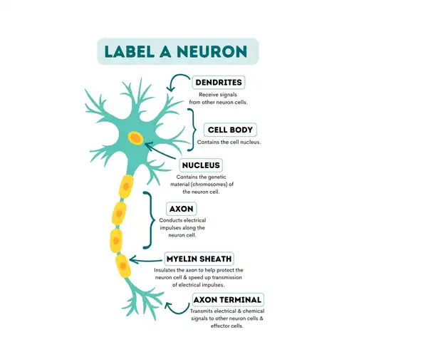
What are the 6 parts of neurons?

- Dendrites: Receive signals from other neuron cells.
- Cell body: Contains the cell nucleus.
- Nucleus: Contains the neuron cell’s genetic material (chromosomes).
- Axon: Conducts electrical impulses along the neuron cell.
- Myelin sheath: Insulates the axon to help protect the neuron cell & speed up the transmission of electrical impulses.
- Axon terminal: Transmits electrical & chemical signals to other neuron cells & effect cells.
Tips on how to label a neuron
There are a few things you should keep in mind when Neuron labeling.
- Make sure to use the correct terminology for the cell type you are labeling.
- Use modern and standardized nomenclature when labeling cells.
- Use arrows to indicate the direction of axons and dendrites.
- Use color coding to make it easy to remember which cell types you are labeling.
- Label cells according to the information they contain, not just their appearance.
- Label cells before you start neuroanatomical reconstruction. This will help you keep track of your progress.
- Label cells accurately and consistently so that your results are reproducible.
- Be patient – Neuron labeling is time-consuming, but it is well worth the effort!
How to label a neuron step-by-step
When Neuron labeling, it’s important to follow three basic steps:
- To identify the cell body, start by marking the center of the image using your cursor. This will help you locate the cell body when you go back and reference it later on.
- To identify the axon, draw an arrow from the center of the image toward the tip of the axon. Make sure to include both the cell body and the axon in your drawing.
- To identify the terminal node, mark where the axon meets the dendrite.
How to label a neuron using the best practices
There are a few different ways to label a neuron, but using a neuron chart is the most accurate and efficient way. A neuron chart is a graphical representation of brain cells, making labeling neurons easy and precise. A neuron chart can be made with paper or online and customized to fit your needs.
To create a neuron chart, start by clustering all the cells in your image into groups according to their function. For example, in the image above, all the red cells are likely associated with blood vessel growth and development, while the green cells may be associated with nerve cell growth and function.
Once you have grouped all the cells, it’s time to label each cell with its corresponding function. To do this, start by drawing a line between each cell and then labeling the line with the name of the cell it connects to (in this example, the red line connects the red cell to the green cell so that we would label this line “red-green”). Be sure to use proper capitalization and punctuation!
Next, it’s important to ensure that the labels are consistent throughout the chart. For example, you don’t want two cells that connect to have different labels (in this example, we would want “red-green” to be the same for both cells). Finally, please take pictures of your neuron chart so you can reference it later (more on taking pictures in a bit!).
What are the different types of neuron labeling approaches?
One of the main ways researchers label neurons is by using different types of markers. Several markers can label neurons, including fluorescent dyes, antibodies, and proteins. Each has its advantages and disadvantages. Here is a recap of the most common types of neuron labels:
- Fluorescent dyes are mainly used to stain neurons for later observation or study. They tend to be more specific and less likely to cross with other cells or tissues.
- Antibodies are proteins that bind to certain molecules in other cells or organisms. By labeling specific antibodies, researchers can track the movement of molecules between cells.
- Proteins: Proteins are complex molecules that play a major role in cellular functions. By labeling proteins, researchers can track their movements and interactions in the cell.
What are the benefits of neuron labeling?
One of the main benefits of neuron labeling is that it can help scientists understand how diseases affect neurons. For example, when scientists study Huntington’s disease, neuron labeling can better understand how the disease progresses and affects different brain parts. By looking at the behavior of individual neurons, they can also develop new methods for treating the disease.
Another benefit of neuron labeling is that it can help scientists study how genes influence neuron function. For example, scientists can learn more about how those genes are involved in motor function, memory formation, and other processes by studying the expression of specific genes in neurons. This information can then be used to develop new treatments or therapies for conditions such as Parkinson’s disease or autism.
Neuron labeling also can be used to study how brain development occurs. By examining the different types of neurons in different stages of development, scientists can learn more about how the brain develops and changes over time. This information is valuable not only for researchers studying neurodegenerative diseases but also for developmental psychologists who study learning and memory.
In addition to these benefits, neuron labeling is relatively safe. Most techniques used to label neurons are non-invasive and do not require any special equipment or expertise. However, there are a few risks associated with neuron labeling. For example, if the label is not properly applied, it could damage the cells or lead to inaccurate results.
Additionally, some neuronal proteins are difficult to detect and may not be visible with traditional labeling methods. Finally, some labeling techniques can have unintended consequences in cells or animal models, such as causing genetic mutations or changes in brain function.
Neurological labeling is an important tool that can help scientists learn much about how neurons function and how diseases affect them. By carefully choosing which approach to use, scientists can ensure that their research is accurate and informative.
What are the risks of neuron labeling?
There are a variety of risks associated with neuron labeling. Some of the most common include:
- Confusion and inaccuracies in cell or animal data
- Potential side effects, including death
- Risks associated with the methods used to label neurons (e.g., radiation exposure, toxic chemicals)
It’s important to be aware of these risks before you label neurons, so you can make informed decisions about whether or not to proceed with the procedure. If you choose to label neurons, be sure to consider all of these precautions!
Read Also: 50 Amazing Binder Spine Label Template, Benefits, Tips & Steps to Create It
Where are neurons located in the brain?
Neurons are located in the brain. You’ve probably heard this before, but let’s be clear: Neurons are located in the brain. And they can be labeled using antibodies.
As it turns out, there is a lot of interest in understanding how neurons work and their role in our cognitive processes. By labeling neurons, researchers can better understand how different parts of the brain communicate with each other. This knowledge could lead to new treatments for Alzheimer’s and autism spectrum disorder (ASD).
Labeling neurons also have practical applications outside research labs; for example, doctors use antibodies to identify and diagnose infections by identifying specific proteins on cells that form part of an infection’s fingerprint.
Antibodies have many other uses, too – from monitoring environmental pollutants to diagnosing food allergies. So not only does labeling neurons provide insights into neuroscience, but it also has far-reaching impacts across multiple industries and disciplines!
Conclusion
Neurons are one of the key building blocks of the brain, and understanding their function is essential to understanding how the brain works. This blog post discussed where neurons are located in the brain and tips on how to label a neuron. I hope you found this information helpful!